
973-998-8309
Affiliates of
New Jersey Sports Medicine Office Hours:
Monday-Friday 8:30AM - 4:30PM | Phone: 973-998-8301
Phalangeal Fractures
Jay E Bowen, DO, Chief Editor: Craig C Young, MD, Additional Contributor: Gerard A Malanga, MD, Alice Tzeng, MD, Elena Napolitano, MD, Acknowledgements: Andrew D Perron, MD
Practice Essentials
Hand injuries are very common in all sports, especially in ball-playing athletes. Most athletic hand injuries are closed hand injuries and include ligamentous injuries, fractures and fracture-dislocations, tendon injuries, and neurovascular problems. There is increasing recognition that fractures and dislocations of the hand can result in long-term pain and disability if they are not recognized and treated early. [1, 2, 3, 4, 5]
Extra-articular fractures of the distal phalanx are common and are associated with significant soft-tissue injury. Most distal phalangeal fractures are crush injuries from a perpendicular force. They can be associated with significant debility, usually in the form of soft-tissue loss, nail bed injury, or posttraumatic neuromas. Intra-articular fractures of the distal phalanx can result from avulsion of either the extensor tendon, also known as mallet fractures, or of the flexor digitorum profundus, also known as jersey fractures. These can be associated with either small dorsal fragments or larger articular fragments with volar subluxation of the volar fragment. Conservative management is usually the standard of treatment.
Fractures of the proximal phalanx are more common than fractures of the middle phalanges. Dorsal or palmar angulation may occur with these fractures, depending on their location. Nondisplaced fractures are usually stable and are treated with closed reduction and fixation. [2, 6] If significant comminution or segmental bone loss is present, these unstable fractures may require either internal or external fixation.
The proximal interphalangeal (PIP) joint is particularly vulnerable to injury as either an ligamentous or intra-articular fracture, with or without subluxation or dislocation. Middle phalangeal articular fractures at the PIP joint include dorsal lip fractures, palmar lip fractures, and central articular disruptions or pilon fractures. Avulsion and impaction sheer are 2 fracture mechanisms.
Middle phalanx palmar lip fractures are the most common form of osseous injury associated with PIP joint fracture-dislocations. Dorsal fracture-dislocation of the PIP joint is reported to occur in 9 of every 100,000 people each year. Many of these injuries are frequently ignored or treated inappropriately. As a result, there can be permanent swelling, pain, and variable degrees of stiffness, angulation, and degenerative changes.
If a serious phalangeal injury is suspected, radiographs should be performed before more forceful testing. Hand fractures in the athlete are treated with adequate alignment, immobilization, and then motion. In general, intra-articular fractures must be reduced anatomically.
Reduction requires early recognition of the exact location of the fracture and having a complete understanding of the muscle pull on the fragments, then minimizing the deforming force.
See the image below.
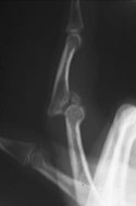
Acute dorsal proximal interphalangeal joint fracture-dislocation.
Functional Anatomy
The phalanges do not contain muscle bellies, and motor function is accomplished only by the flexor and extensor tendons. An overview of the muscles and tendons of the hand is necessary. The thenar muscles consist of 3 intrinsic muscles including the abductor pollicis brevis (which abducts the thumb), the flexor pollicis brevis (which flexes the proximal phalanx of the thumb), and the opponens pollicis (which produces opposition of the thumb).
All 3 intrinsic thenar muscles are supplied by the recurrent branch of the median nerve. The adductor pollicis adducts the thumb and is supplied by the deep branch of the ulnar nerve. The hypothenar muscles are also supplied by the deep branch of the ulnar nerve.
The abductor digiti minimi abducts the fifth digit and flexes its proximal phalanx. The flexor digiti minimi is deeper and also flexes the proximal phalanx of the fifth digit. The opponens digiti minimi, as its name implies, opposes the fifth digit.
The lumbricals are 4 muscles that arise from the tendons of flexor digitorum profundus. Their tendons insert into the radial side of each of the proximal phalanges of the fingers and into the dorsal hood. They flex the metacarpophalangeal joints and extend the interphalangeal joints. The first and second lumbricals are supplied by the median nerve, and the third and fourth lumbricals are supplied by the ulnar nerve.
The palmar and dorsal interossei arise from the metacarpals. The palmar interossei insert into the proximal phalanx and the expansion of the extensor digitorum communis. The palmar interossei are adductor muscles. Dorsal interossei are abductors and insert into the proximal phalanges and the dorsal digital hood. The interosseous muscles are all supplied by the deep branch of the ulnar nerve.
As the tendons of the long flexor and extensor muscles reach the hand, the flexor tendons must first pass deep to the flexor retinaculum and the extensor tendons must pass under the extensor retinaculum. Flexor tendons on the palmar side are anchored to the phalanges by fibrous flexor sheaths to prevent "bow-stringing." Synovial sheaths prevent friction from occurring between fibrous flexor sheaths and the tendons. Synovial sheaths are present on the dorsum of the hand deep to the extensor retinaculum. They extend from a point proximal to the retinaculum to a point in the proximal one third of the dorsum of the hand.
Anatomy of the distal interphalangeal (DIP) joint includes the insertion of the extensor tendon on the distal phalanx.
Sport-Specific Biomechanics
The PIP joint is the most commonly injured area in the hand. There is both anatomic and functional complexity to this joint, which consists of the articulation of the proximal end of the middle phalanx and the distal end of the proximal phalanx. It is a hinge joint with range of motion from 0 º to 120 º in the extension-flexion plane, with the bulk of static and dynamic stability provided by the surrounding ligaments and tendons.
The capsule surrounding the articular surface is composed of the volar plate, thick collateral ligaments, and the extensor tendon dorsally, which divides into 3 slips as it passes over the proximal phalanx. The central slip of the extensor tendon passes directly over the joint and inserts on the dorsal base of the middle phalanx. The lateral bands of the extensor tendon combine distally with the tendons of the intrinsic hand muscles (the retinacular ligaments) to form the extensor tendon that attach to the distal phalanx.
The thick ulnar and radial collateral ligaments of the PIP joint combine with the volar plate to provide lateral stability. The volar plate, a thick fibrocartilaginous structure, forms a sturdy attachment to the middle phalanx where it becomes continuous with the articular cartilage. This limits extension of the PIP joint beyond 0 º.
Proximally, the volar plate forms a thin continuous attachment with the synovial reflection. The lateral margins remain thick strong ligaments. This results in a cul-de-sac between the proximal half of the volar plate and the head of the proximal phalanx, which allows the base of the middle phalanx to glide along the articular surface of the proximal phalanx as the finger flexes. Thus, the volar plate becomes both a static stabilizer limiting hyperextension beyond 0 º and a dynamic stabilizer that influences the position of the flexor tendons at initiation of PIP joint flexion.
History
Distal phalangeal fractures
- Most distal phalangeal fractures are crush injuries from a perpendicular force, as in injuries from a car door or hammer or in sports when a player has a digit stepped on or crushed between the helmets of opposing players. Tuft fractures are often comminuted and are generally stable fractures because of intrinsic splinting of bony fragments by fibrous septae in the fingertips.
- Physical examination in a person suspected of having a phalangeal fracture starts with inspection, attitude of the injured finger, and localization of any swelling. Neurovascular status should be examined as well as color, capillary refill, and digital temperature.
- Palpation of the joint over 4 planes (ie, dorsal, volar, medial, lateral) allows assessment of point tenderness over ligamentous origins and insertions, which is suggestive of soft-tissue disruption.
- Passive range of motion and joint stability should be assessed through dorsal, volar, and lateral stressing. It should not be assumed that lack of full active flexion or extension is merely secondary to joint pain.
- Fractures at the base of the distal phalanx are usually mallet avulsion fractures and are caused by rupture of the extensor tendon at the DIP joint. These are common injuries in basketball or baseball players. [7] The mechanism of injury usually results from a direct blow to the tip of the finger that causes forced flexion of the DIP joint.
Jersey finger
- Avulsion of the flexor digitorum profundus from its insertion on the volar base of the distal phalanx results in the jersey finger. The mechanism of injury most commonly occurs in football when a player grabs the opponent's jersey. There is forced hyperextension of the DIP joint against an actively contracting flexor tendon. The ring finger is most commonly involved. Pain and swelling of the affected finger occurs, with loss of active flexion of the DIP joint.
- This injury is often overlooked because on physical examination there is no obvious deformity present and the loss of DIP flexion is either not appreciated or dismissed secondary to pain or soft-tissue swelling. If one suspects flexor tendon avulsion, then gentle active flexion of the DIP joint should be tested specifically. Radiographic findings are often normal but may reveal a small avulsion fracture.
Middle phalangeal fractures
- The mechanism of injury of middle phalangeal fractures is usually the result of a blunt or crush force perpendicular to the long axis of the bone. Angulation and rotation are 2 features of instability that must be examined.
- Visual inspection usually detects dislocations and subluxations but is most valuable in the angulatory or rotary deformities that can accompany subluxation/dislocations. These deformities suggest complications ranging from asymmetric ligament tears to interposed tissues between joint surfaces. Rotational deformities are serious injuries and are detected clinically. Patients should be asked to fully flex the phalanges, and the long axis of the fingers should point to the scaphoid tubercle or the distal radius with the fingers parallel to each other.
- Dorsal or volar angulation is evaluated radiographically in relation to the insertion of the flexor digitorum superficialis. Fractures distal to the tendon result in volar angulation, and proximal fractures result in dorsal angulation from the resulting muscle pull.
Proximal phalangeal fractures
- Proximal phalangeal fractures are relatively common and may result in a great deal of disability. The mechanism of action usually results from a direct perpendicular force, a rotary force, or hyperextension of the finger.
- The physical examination is the same as that for distal phalangeal fractures (see above).
Dorsal PIP joint dislocations
- Dorsal dislocation of the PIP joint is a common injury that occurs secondary to joint hyperextension. The volar plate must be ruptured for dorsal dislocation to occur. This rupture usually occurs at the distal insertion site at the base of the middle phalanx and may involve a tiny avulsion fracture of the base of the middle phalanx.
- When disruption of the collateral ligaments has also occurred, rotary deformity and lateral stress instability will be apparent. Joint dislocation should be assessed for collateral ligament injury, because any associated collateral ligament problems require orthopedic referral.
Volar PIP joint dislocations
- Volar dislocations of the PIP joint are less common than dorsal dislocations and are more difficult to manage than other injuries. A volar dislocation ruptures the extensor mechanism, possibly involving injury of the central slip.
- Evaluation of the PIP joint injury starts with the history to understand the mechanism and direction of injury, the presence of an initial deformity, and whether a reduction was performed. The physical examination includes thorough inspection and palpation of the area and localization of the tenderness. The examination should also include testing active and passive range of motion, with and without resistance. Tenderness dorsally indicates a central slip injury. This is confirmed if there is pain and inability to extend the PIP joint against resistance. Lateral stressing of the joint assesses integrity of the collateral ligaments and the degree of instability.
Grading and treatment
-
Instability of the PIP joint is classified as 3 grades, and treatment is based on the grading of the injury. Grading is based on active range of motion and stress testing. When pain interferes with evaluating for these, a local anesthetic block at the metacarpal level should be performed.
- Grade I injuries are stable through an active range of motion and do not angulate more than 20° greater than the contralateral side on stress testing.
- Grade II injuries will not deform with active range of motion, but stress testing will show instability, defined as greater than 20° of angulation when compared with the contralateral side. These injuries are due to a complete disruption of one of the major retaining ligaments, which include the radial or ulnar collateral ligaments, the volar plate, or the central extensor slip.
- Grade III injuries show deformity with active range of motion or the injured fingers are unable to be moved through more than 75% of the range of motion under anesthetic block. This implies complete disruption of greater than 50% of the entire capsule or 2 or more of the major retaining ligaments.
- Collateral ligament injuries may be the result of a pure angulatory stress or a joint dislocation with the radial collateral ligament as the ligament most frequently injured.
The most common complication of a volar plate injury or dorsal dislocation is the development of pseudo-boutonniere deformity, which is a persistent PIP joint flexion contracture. Another common problem is detachment of the volar plate, resulting in hyperextension laxity at the PIP joint and a swan-neck deformity. Associated with the swan-neck deformity is the painful snapping of the lateral bands with attempted flexion. Both of these complications can be prevented with appropriate treatment.
DIP joint dislocations: Dislocations of the DIP joint are uncommon. Such injuries are usually the result of a hyperextension force, and they are frequently open because the skin in this area is both thin and well anchored.
Boutonniere deformity
- Disruption of the central slip of the extensor digitorum communis tendon over the PIP joint produces the classic boutonniere deformity. This can be caused by blunt trauma over the dorsal aspect of the finger, with a deep laceration over the PIP joint, or with forced flexion of the joint.
- Avulsion of the central slip results in a flexion deformity of approximately 15-30° due to unopposed pull of the flexor digitorum superficialis tendon. As a result of loss of extensor tendon function, in addition to localized pain and swelling, the PIP joint cannot be actively extended. One can think of this as the PIP "buttonholing" through the central slip.
- Prevention of a boutonniere deformity requires a high index of suspicion during the acute stage of the injury. A central slip injury should be suspected in patients who present with pain mainly over the dorsal aspect of the PIP joint.
- Radiographic findings are rare for the small avulsion fracture at the dorsal base of the middle phalanx. The best indication of central slip damage is inability to extend the PIP joint fully against active resistance. A central slip disruption is present unless proven otherwise when full extension cannot be performed.
- Immediately after injury, examination will reveal a diffusely swollen PIP joint held in mild flexion. Active extension may be possible with the lateral bands. The diagnosis is made by palpating all 4 quadrants of the joint. This is confirmed if there is pain and inability to extend the PIP joint against resistance. Tenderness dorsally indicates a central slip injury.
- Boutonniere deformities are graded I through IV.
-
- A grade I injury is correctable passively.
- A grade II injury is the case in which there is a PIP joint flexion contracture of less than 30° that is not passively correctable.
- Grade III boutonnieres demonstrate a PIP joint flexion contracture greater than 30° and loss of flexion of the distal joint.
Imaging Studies
If a serious injury is suspected, radiographs should be performed before more forceful testing.
- Most hand fractures are usually detected by obtaining 3 views (ie, anteroposterior [AP], a true lateral, oblique) of the specific injured joint rather than the entire hand.
- Brewerton views (beam angled 30° from the ulnar side of the hand) can be used to detect collateral ligament avulsion injuries.
- Direct posterior-anterior (PA) and lateral views of the thumb should be obtained if the thumb is suspected of injury. Fractures of the middle and proximal phalanx may angulate palmar or dorsally.
- Postreduction radiographs should show no more than 10° of angulation and rotational displacement.
When a patient presents with a dorsal dislocation of the PIP joint, a prereduction radiograph should be obtained to rule out any associating fracture, which could interfere with the attempted reduction.
Procedures
If a patient with a suspected phalangeal fracture is having significant discomfort that cannot allow adequate testing, anesthesia with a digital nerve block should be performed.
Remember to perform a sensory nerve examination before administering the nerve block.
Fractures of the distal phalanx
Tuft fractures are treated by caring for the accompanying soft-tissue injury and splinting of the finger to prevent further discomfort or injury. A variety of splinting devices can be used for loose protection. In the closed crush fracture of the distal phalanx, the L -shaped Alumafoam splint placed on the volar aspect to protect the soft tissues is considered the best treatment. Tight circumferential taping around the fingertip should not be used because of an increased risk of circulatory compromise. Splinting is generally maintained for about 2-3 weeks.
Avulsion of the nail plate and injury to the nail bed is often associated with tuft fractures. It is necessary to reduce the nail plate under the eponychium, and if this cannot be performed, the plate can be removed. The distal phalanx may be destabilized to some extent, but as there are no tendons attached to the tuft of the distal phalanx, these injuries rarely displace. If the nail is removed during repair, packing of the eponychial space with petroleum gauze is used to prevent scarring and closure of the space, which could lead to stunted nail growth. Tuft fractures may progress to nonunion but are usually asymptomatic.
Open fractures of the distal phalanx require thorough cleansing, debridement, and inspection for foreign bodies. Orthopedic assistance is not required for uncomplicated closures. Open fractures with extensive soft-tissue damage are frequently associated with chronic pain and disability and may need orthopedic assistance. Open fractures of the distal phalanx require a course of antibiotic treatment.
The shaft of the distal phalanx is very narrow and mostly cortical. Fractures at this location can be problematic. Nonunion can be symptomatic; occasionally, these require internal fixation.
Mallet finger
Acute phase
The mallet finger is a stable injury. If the patient presents with lack of active extension of the DIP joint, then splinting of the finger in extension or slight hyperextension for 8 weeks is the treatment of choice. It is important to educate these patients not to remove the splint. Observed these patients in the office every 1-2 weeks to ensure that the splint is not being removed.
Patients can be instructed to change the splint every few days to allow cleansing of the skin. However, reapplication of the splint should always be completed with assistance. The DIP joint should not be allowed to flex at any time. If the tendon ruptures from DIP flexion, then the entire 8-week period of splinting must be repeated.
When splinting the mallet finger, the DIP joint should not be severely hyperextended, as the skin over the dorsum of the joint will blanch and slough, and ischemia and necrosis of the dorsal tissue may occur. There are cases in which a patient cannot tolerate an external device, and these patients are treated with transarticular Kirschner wire (K-wire) fixation for 6-8 weeks. The PIP joint should be in full motion, because splinting of the PIP may result in thickening of the collateral ligaments and subsequent joint contracture and stiffness.
Rehabilitation is much longer and more difficult with a stiff PIP and mallet finger. However, it should be noted that the PIP joint is infrequently immobilized in the hyperflexible patient to allow the terminal tendon to heal in a shorter position.
Maintenance phase
Full activity is allowed during the 8-week interval of continuous splinting for a mallet finger. However, additional finger protection and supervision by an athletic trainer or physician is required during contact sports. The splint may be removed during the day after 8 weeks. Tendon stability is maximized if the splint is used at night and during athletic activity for another 8 weeks.
Patients with an undiagnosed or neglected mallet finger may undergo a trial of prolonged splinting for up to 3 months; however, the prognosis is best when a splint is applied within 2 weeks of the injury. Surgical fusion of the DIP joint can provide stability if splinting fails in these cases.
Surgical intervention
The mallet finger is a stable injury. However, treatment is more difficult if a portion of the bone is avulsed from the distal phalanx. If the bone can be reduced closed in an adequate position with less than 2 mm of displacement, then closed treatment is recommended.
Jersey finger
Acute phase/surgical consultation
See the list below:
- A type 1 jersey finger injury is an avulsion of the flexor digitorum profundus tendon into the palm, losing its blood supply from both the bone and the vincular system. These injuries require reattachment within 7-10 days to avoid necrosis of the tendon.
- In type 2 injuries, the tendon retracts to the level of the PIP joint and is held by an intact vincula. A reattachment can be successful anywhere from 3 to 6 weeks after injury.
- Type 3 injuries involve avulsion of a large fragment of bone from the palmar base of the distal phalanx. The tendon does not retract past the DIP joint because of the fracture fragment being caught by the A4 pulley. Type 3 injuries are not to be treated with closed reduction because of the possibility of an associated avulsion of the profundus tendon from the osseous fragment. Delayed repair also can be successful.
Fractures of the proximal phalanx
Acute phase
Dynamic splinting is the treatment of choice for nondisplaced fractures. Fractures of the shaft usually result in volar angulation, because the intrinsic tendons pass obliquely from a proximal volar to a distal dorsal direction. Angulation is also increased by the influence of the extensors and flexors compressing bone longitudinally. Reduction is accomplished by longitudinal traction and flexion of the metacarpophalangeal joint to lessen the pull of the intrinsic muscles and then followed by longitudinal traction and flexion of the distal fragment.
Maintenance phase
For displaced fractures, splinting is used after reduction, with the wrist in slight extension and the metacarpophalangeal joint flexed to 70°. Free motion is allowed at the PIP and DIP joints for early tendon motion. Oblique and spiral fractures are usually unstable after reduction and require splinting with either ulnar or radial gutter splints that extend to the distal phalanx. In the proximal phalanx, the surrounding tendons lie in close proximity to the periosteum, and there is a susceptibility to adhesions and stiffness during the healing process. Therefore, appropriate fracture alignment, stability, and early motion are very important. [8] One study in patients with extra-articular fractures of the proximal phalanges examined the use of a cast that allowed unrestricted motion of the wrist, and that fixed the MCP joints in 70 to 90 degrees of flexion with buddy taping of the fingers. [9] This cast allowed quicker return of wrist motion and showed similar healing of fractures compared toastatic forearm-fingercastinan intrinsic-plus position. [9]
Fok et al report on 10-year results managing proximal phalangeal fractures with dynamic treatment. [10] A dynamic splint that kept the metacarpophalangeal joint maximally flexed while allowing free movement of the proximal and distal interphalangeal joints of the injured finger was applied for at least 4 weeks. Results were evaluated using the Belsky classification and grip strength assessment. The results of 97 patients (103 fractures) were analyzed. At a minimum 1-year follow-up, 75% of patients attained excellent or good results. Neither nonunion nor delayed fracture union was noted. The 25% of patients who attained poor results were older than those who attained excellent or good results (average age, 53.1 vs 35.1 years, respectively) and tended to comply poorly with the rehabilitation program. Using the stabilizing effect of the zancolli complex-metacarpophalangeal retention apparatus and a metacarpophalangeal block splint, bone healing and movement recovery can be achieved simultaneously. [10]
PIP fracture dislocations
Posterior PIP fracture-dislocations occur with volar lip fractures and volar dislocations with dorsal avulsion fractures. Posterior fracture-dislocations usually involve some degree of axial compression, comminution, and involvement of the volar articular surface. Treatment methods are grouped into the following 5 categories: static immobilization, dorsal extension block splinting, longitudinal traction, open reduction and internal fixation, and palmar plate arthroplasty.
Acute phase
A splint or transarticular K-wire can be used for static immobilization. Articular reduction must be monitored serially by radiographs, and immobilization for more than 3 weeks may result in stiffness. The PIP joint must be immobilized in about 30° of flexion for about 2 weeks.
Maintenance phase
PIP motion is initiated, but significant hyperextension of the joint may injure healing tissues. An additional extension block splint prevents the final 30° of extension while allowing joint flexion. The ideal case for extension block splinting is when there is some collateral ligament restraint remaining on the major fragment. Therefore, a fracture involving less than 40% of the articular surface is preferred for extension block splinting. Lateral radiographs must be monitored to ensure that subtle subluxation is not present and that dorsal joint surfaces are reduced adequately.
Rehabilitation phase
At 6 weeks, passive flexion and strengthening should be instituted. As soon as the splint is off, grip and massage exercises and functions are started. Avoid pinching of the involved finger and stretching of the involved joint. When grip strength reaches the 50% level, then full freedom of function is given. When the patient is fully asymptomatic, full PIP motion is encouraged with protective splinting used during athletic activity. Formal rehabilitation is usually discontinued at 3 months after injury.
Palmar lip fractures
Acute phase
Stable palmar lip fractures (< 30% of the articular surface) do not cause PIP joint subluxation and are treated by programs that maximize motion. An extension block splint may be used for stable hyperextensible palmar lip fractures or a double Alumafoam splint method to achieve 20° of PIP joint flexion for 3 weeks; then, it is adjusted to 10° of additional extension over the next 2 weeks.
Rehabilitation program
For pilon fractures, traction is discontinued after 6 weeks, and aggressive range of motion is maintained.
S
urgical Intervention
Mallet fingers involving fractures of more than 25-30% of the articular surface, or if volar subluxation of the DIP joint is present, then open reduction and internal fixation is indicated. Fixation is continued for 4-6 weeks, after which the pins are removed and motion is begun. The patient wears an external splint for the month after internal fixation is discontinued.
For jersey fingers, with continued extension vector force, the distal phalanx continues into hyperextension. Subsequently, a secondary intra-articular fracture at the base of the distal phalanx occurs as the distal portion of the middle phalanx is driven into the base of the distal phalanx. Avulsion of the flexor digitorum profundus with a separate intra-articular fracture of the distal phalanx has been proposed as a type 4 injury. Treatment consists of open reduction and internal fixation of the intra-articular fracture with reinsertion of the profundus tendon.
For fractures of the middle phalanx, if conservative treatment is not sufficient, then percutaneous pinning or open reduction using K-wires is used. [11] Plates or screws are not regularly used at the phalangeal level because there is too much soft-tissue damage in the process (although Giesen et al reported successful treatment of proximal and middle phalangeal fractures in 26 patients with intramedullary headless compression screws [12] ). Displacement of the bone around the center of the longitudinal axis is rotational.
Malrotation is detected clinically, not radiographically. Patients should be asked to fully flex the phalanges; the long axis of the fingers should point to the scaphoid tubercle or the distal radius with the fingers parallel to each other. If suspected, a rotational deformity warrants surgical referral. A rotational deformity is often associated with spiral, oblique, or comminuted fractures.
A comminuted fracture of the base involving the PIP joint can be difficult to treat. In these cases, traction with a transverse K-wire through the middle or distal phalanx can be used. They are removed after 6 weeks, but external splint immobilization is needed for an additional 2-4 weeks.
An alternative method, as described by Eaton, is primary volar plate arthroplasty in which the comminuted volar portion of the middle phalanx is removed and the volar plate is advanced into the defect.
For PIP fracture-dislocations, if a congruent reduction cannot be maintained or if more than 30-50% of the articular surface is involved, then arthroplasty may be required. If there is a large volar fragment, then internal fixation may be necessary. It is believed that fragments with greater than 2 mm of displacement lead to excessive extensor deficit and, therefore, warrant open reduction and internal fixation.
Successful surgical treatment of PIP fracture-dislocations is dependent on the following principles. The first is to reestablish the normal flexion glide of the middle phalanx around the proximal phalanx head during the flexion arc. Hinging at the fracture site must be avoided. The surgeon must eliminate joint subluxation and then reestablish joint stability. Second, early motion is initiated whenever possible to enhance cartilage and soft-tissue healing and also to minimize adhesions or contractures. Anatomic restoration of the fractured joint surface is desirable but is a much less important treatment goal.
The preferred treatment for unstable palmar lip fractures is palmar plate arthroplasty, and referral to a surgeon is necessary. Pilon fractures also require a surgical consult. Pilon fractures are placed in traction, and patients should begin active and passive motion as soon as possible. Percutaneous pinning or limited open reduction and internal fixation may be necessary.
A prospective, longitudinal study by Miller et al found that in patients who underwent open reduction and internal fixation of proximal phalangeal fractures, with rehabilitation begun within 1 week of fixation, the greatest degree of recovery with regard to range of motion, hand use, pain, strength, and work participation was seen by the sixth week postoperatively. The study involved 66 patients. [13]
Other Treatment
Dorsal PIP joint dislocations
A dorsal PIP joint dislocation is reduced easily with traction on the finger, followed with flexion of the PIP joint. After reduction, joint stability is provided by the intact collateral ligaments. As long as the reduction is stable, the joint congruent, and the fragment, if present, is small and minimally displaced, a dorsal extension block splint is used. Large displaced fragments lead to joint instability and respond best to operative treatment. However, some comminuted fractures can be treated only by traction and early range of motion. No treatment is likely to provide complete pain-free range of motion in these injuries.
The PIP joint should be immobilized in approximately 30° of flexion for 2-3 weeks. Buddy taping or other protective splinting should be used for another 3-4 weeks during activity or sports participation. The most important principle is the prevention of hyperextension, which could reinjure the volar plate.
If the dorsal PIP joint dislocation cannot be reduced because the proximal phalangeal head is impinged between the central slip and the lateral bands, then open reduction is required. Otherwise, these injuries can be treated with buddy taping, which is worn continuously for the first 3 weeks and then only during physical activities for an additional 4-6 weeks. Complete resolution of pain usually takes 4-6 months, although a slight residual swelling is often permanent.
Grade I and II injuries are treated with extension block splints, which limit the last 20-30° of extension but allow full flexion of the distal joint. Active protected extension is begun at 2 weeks, and athletes can be allowed to play with buddy taping. Protection should be continued for 6-8 weeks or until joint motion is pain free.
Grade III injuries, which includes irreducible dislocations, usually require surgery. If left undertreated, grade III injures can lead to permanent deformity, lost motion, and degenerative joint changes. Radiographs should be taken in the AP, lateral, and oblique planes.
Volar PIP joint dislocations
As with dorsal dislocations, the head of the proximal phalanx may become impinged between the central slip and the lateral band, in which case open reduction is necessary. Another indication for open repair is if the joint is functionally unstable (ie, the joint dislocates spontaneously as the patient moves it). A third indication for open reduction is an untreated chronic volar plate avulsion that allows the joint to hyperextend.
After closed reduction of a volar dislocation, the PIP joint should be immobilized in extension for 6-8 weeks.
All patients with closed injuries of the PIP joint should be told that swelling and disability are common for possibly months, with swelling and disability persisting for 6 months to 1 year in some complete ligamentous injuries. In addition, some thickening is likely to be permanent.
Collateral ligament injuries
Grade I or II injuries may be treated by immobilization in 15-20° of flexion for 2-3 weeks, followed by buddy taping for 3-6 weeks or until symptoms subside. Resumption of athletic activities is possible with these injuries, but reinjury is common, with risk of a grade III injury. A radiograph showing joint symmetry and stability should be obtained before administering this treatment.
DIP joint dislocations
If the dislocation is closed, it should be reduced after anesthetizing the digit by applying longitudinal traction. After reduction, the joint should be evaluated for tendon and ligament injuries. They can be irreducible when a condyle of the middle phalanx becomes buttonholed between a collateral ligament and the volar plate.
Treatment consists of digital block anesthesia followed by one gentle attempt at closed reduction by hyperextending the distal phalanx and then sliding the base of the distal phalanx over the head of the proximal phalanx. Rotational force may be necessary. If unsuccessful, open reduction is required.
Open DIP joint dislocations require surgical wound care and reduction to prevent bone or joint infections, even if there is only a small break in the volar skin. Otherwise, the distal joint is splinted in full extension for 1-2 weeks. Active range of motion is begun at 2-3 weeks, and a splint is worn until full, pain-free range of motion is achieved. If the athlete continues to play sports, the DIP joint should be splinted for 4-6 weeks to prevent reinjury.
Boutonniere deformity
Both grade I and II injuries are treated by splinting the PIP joint in full extension, leaving the distal joint free to actively flex. The adjacent metacarpophalangeal and DIP joints should be allowed to have full range of motion. Splinting or buddy taping should continue for 6-8 weeks until there is pain-free motion. During this time, a program of active and passive range of motion at the DIP joint remobilizes the lateral bands and allows the central slip to heal at its proper length.
Grade III boutonnieres demonstrate a PIP joint flexion contracture greater than 30° and loss of flexion of the distal joint. An effort is made to correct the PIP flexion contracture by splinting or casting before an operative procedure that includes release of any residual contracture.
Grade IV deformities present with a fixed PIP flexion contracture and degenerative change in the joint. For treatment, the PIP joint is held in hyperextension. Surgery results in little gain of active motion.
Arthrodesis is often necessary to correct this deformity. Patients with a chronic boutonniere deformity, either from misdiagnosis or neglect, should be referred to a hand surgeon for evaluation and treatment. Complete recovery may not be possible at this time.
Medication Summary
Pain control is the mainstay of treatment in patients with phalangeal fractures.
Analgesics
Class Summary
Pain control is essential to quality patient care. Analgesics ensure patient comfort, promote pulmonary toilet, and have sedating properties, which are beneficial for patients who have sustained trauma or injuries.
Acetaminophen and Codeine (Tylenol #3)
Indicated for the treatment of mild to moderate pain.
Hydrocodone bitartrate and acetaminophen (Vicodin ES, Lortab, Lorcet, Norcet)
Drug combination indicated for moderate to severe pain.
Antibiotics
Class Summary
Antibiotics are used for open contaminated wounds. Therapy must cover all likely pathogens in the context of this clinical setting.
Cefazolin (Ancef, Kefzol, Zolicef)
First-generation semisynthetic cephalosporin that arrests bacterial cell wall synthesis, inhibiting bacterial growth. Primarily active against skin flora, including Staphylococcus aureus. Typically used alone for skin and skin-structure coverage. IV and IM dosing regimens are similar.
Follow Up
Return to Play
Return to play in patients with phalangeal fractures is guided by the patient's symptoms, healing, and potential for reinjury. If the fracture can be adequately protected and immobilized, while not interfering with the patient's ability to participate, then sports participation can be allowed, providing the patient has adequate pain control.
Complications
Phalangeal fractures, as with all fractures, are subject to the risks of delayed union, malunion, and nonunion. These can be the result of inadequate immobilization and patient noncompliance with immobilization.
Prognosis
Most phalangeal fractures heal without significant complications. Fractures that involve a joint are more prone to prolonged stiffness and decreased range of motion.
Onishi et al conducted a study consisting of 70 patients with 75 unstable proximal phalangeal fractures to determine the risk factors for postoperative finger stiffness after open reduction and internal fixation of unstable proximal phalangeal fractures using a low-profile plate and/or screw system. The study found that plate fixation and dorsal placement were independent risk factors for finger stiffness. The study recommended the use of screw fixation as much as possible for unstable proximal phalangeal fractures using a midlateral approach. [14]
References
- Ruby LK. Common hand injuries in the athlete. Orthop Clin North Am. 1980 Oct. 11(4):819-39. [Medline].
- Hoffman DF, Schaffer TC. Management of common finger injuries. Am Fam Physician. 1991 May. 43(5):1594-607. [Medline].
- Mastey RD, Weiss AP, Akelman E. Primary care of hand and wrist athletic injuries. Clin Sports Med. 1997 Oct. 16(4):705-24. [Medline].
- Aitken S, Court-Brown CM. The epidemiology of sports-related fractures of the hand. Injury. 2008 Dec. 39(12):1377-83. [Medline].
- Kamath JB, Harshvardhan, Naik DM, Bansal A. Current concepts in managing fractures of metacarpal and phalangess. Indian J Plast Surg. 2011 May. 44(2):203-11. [Medline]. [Full Text].
- Belsky MR, Eaton RG, Lane LB. Closed reduction and internal fixation of proximal phalangeal fractures. J Hand Surg [Am]. 1984 Sep. 9(5):725-9. [Medline].
- Wilson RL, McGinty LD. Common hand and wrist injuries in basketball players. Clin Sports Med. 1993 Apr. 12(2):265-91. [Medline].
- Singh J, Jain K, Mruthyunjaya, Ravishankar R. Outcome of closed proximal phalangeal fractures of the hand. Indian J Orthop. 2011 Sep. 45(5):432-8. [Medline]. [Full Text].
- Franz T, von Wartburg U, Schibli-Beer S, Jung FJ, Jandali AR, Calcagni M, et al. Extra-articular fractures of the proximal phalanges of the fingers: a comparison of 2 methods of functional, conservative treatment. J Hand Surg Am. 2012 May. 37(5):889-98. [Medline].
- Fok MW, Ip WY, Fung BK, Chan RK, Chow SP. Ten-year results using a dynamic treatment for proximal phalangeal fractures of the hands. Orthopedics. 2013 Mar. 36(3):e348-52. [Medline].
- Klein DM, Belsole RJ. Percutaneous treatment of carpal, metacarpal, and phalangeal injuries. Clin Orthop Relat Res. 2000 Jun. 375:116-25. [Medline].
- Giesen T, Gazzola R, Poggetti A, Giovanoli P, Calcagni M. Intramedullary headless screw fixation for fractures of the proximal and middle phalanges in the digits of the hand: a review of 31 consecutive fractures. J Hand Surg Eur Vol. 2016 Sep. 41 (7):688-94. [Medline].
- Miller L, Ada L, Crosbie J, Wajon A. Pattern of recovery after open reduction and internal fixation of proximal phalangeal fractures in the finger: a prospective longitudinal study. J Hand Surg Eur Vol. 2016 Oct 3. [Medline].
- Onishi T, Omokawa S, Shimizu T, Fujitani R, Shigematsu K, Tanaka Y. Predictors of Postoperative Finger Stiffness in Unstable Proximal Phalangeal Fractures. Plast Reconstr Surg Glob Open. 2015 Jun. 3 (6):e431. [Medline].
- Bowers AL, Baldwin KD, Sennett BJ. Athletic hand injuries in intercollegiate field hockey players. Med Sci Sports Exerc. 2008 Dec. 40(12):2022-6. [Medline].
- Khalid M, Theivendran K, Cheema M, Rajaratnam V, Deshmukh SC. Biomechanical comparison of pull-out force of unicortical versus bicortical screws in proximal phalanges of the hand: a human cadaveric study. Clin Biomech (Bristol, Avon). 2008 Nov. 23(9):1136-40. [Medline].
- Matzon JL, Cornwall R. A stepwise algorithm for surgical treatment of type II displaced pediatric phalangeal neck fractures. J Hand Surg Am. 2014 Mar. 39(3):467-73. [Medline].
- Held M, Jordaan P, Laubscher M, Singer M, Solomons M. Conservative treatment of fractures of the proximal phalanx: an option even for unstable fracture patterns. Hand Surg. 2013. 18(2):229-34. [Medline].
- Franz T, Jandali AR, Jung FJ, Leclère FM, von Wartburg U, Hug U. Functional-conservative treatment of extra-articular physeal fractures of the proximal phalanges in children and adolescents. Eur J Pediatr Surg. 2013 Aug. 23(4):317-21. [Medline].
New Jersey Regenerative Institute | 197 Ridgedale Avenue, Suite 210, Cedar Knolls,
New Jersey 07927 | 973-998-8309
Quick Links
Links
Privacy Links


Results may vary based on individual’s overall health, lifestyle, severity of the orthopedic condition being treated and responses are not guaranteed. The information on this site is solely for purposes of general patient education, and may not be relied upon as a substitute for professional medical care. Consult your own physician for evaluation and treatment of your specific condition.